2026-05-06
Due to various causes, when repairing the urethral injury, scar tissue forms and contracts, causing the urethral cavity to narrow and even completely close; the internal epithelium becomes thinner, and some of it undergoes metaplasia into stratified squamous epithelium; the lamina propria becomes discontinuous, and the vascular sinus structure of the corpus cavernosum is replaced by a large amount of fibrous connective tissue, and the connective tissue has no obvious boundary with the corpus cavernosum tissue, and the two interweave with each other. These changes result in urethral stenosis. Generally, it is not recommended to perform intermittent catheterization for patients with urethral stenosis.
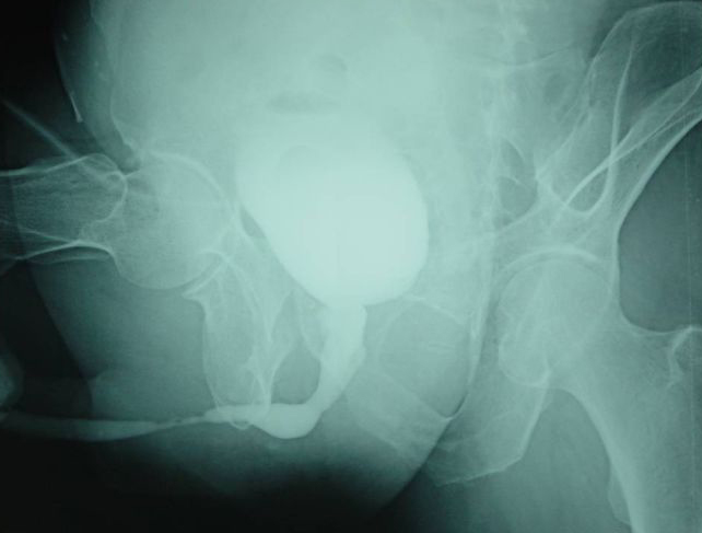
Urethral stricture is one of the common diseases in urology. In recent years, with the development of industries such as transportation and construction, as well as the increase in the use of urethral internal instruments and surgeries, its incidence has been continuously rising in both developed and developing countries. The common causes of urethral stricture include congenital, inflammatory, traumatic, iatrogenic and other unknown causes.
Due to the longer urethra in men compared to women, along with differences in anatomical location and tissue structure, it is more susceptible to factors such as trauma and infection. At the same time, it is also influenced by social factors, and this disease often occurs in adult men.
1. Causes of urethral stricture
Urethral stricture can be classified as spasmodic and organic.
1.1 Spasmodic urethral stricture is a temporary phenomenon, caused by the contraction of the external urethral sphincter. The triggering causes may include urethritis, urethral calculi, the use of urethral instruments, or abnormal sexual desire, etc. Spasmodic stricture can be treated with comprehensive methods, including eliminating the triggers, hot water sitz baths, sedatives and analgesics, and antispasmodic agents, which can usually alleviate the condition.
1.2 Organic stricture can be classified into three types based on the cause:
a: Congenital urethral stricture, such as external urethral orifice stricture, urethral valves, etc.
b: Inflammatory urethral stricture, which can be caused by gonorrhea, tuberculosis, or non-specific infections. The shape of the stricture is more complex and the degree is more severe. Inflammatory urethral stricture is mainly treated with urethral dilation in the early stage after infection control, and the use of indwelling catheters can also cause urethral stricture.
c: Traumatic urethral stricture is the most common type, caused by severe urethral injury, improper initial treatment or untimely treatment.
1.3 The degree, depth and length of the stricture are quite large, and usually there is only one stricture. Gonorrheal stricture may be multiple strictures. The stricture may be secondary to infection, resulting in urethral diverticulum, urethral perineal inflammation, prostatitis or epididymo-orchitis. If the obstruction of the urine flow cannot be relieved for a long time, it may eventually lead to hydronephrosis, renal function damage, and uremia.
2.Clinical manifestations of urethral stricture
The symptoms of urethral stricture can vary depending on its severity, extent and development process. The main symptoms are as follows.
2.1 Difficulty in urination, with severe cases resulting in urinary retention. Initially, urination is difficult, the duration is prolonged, the urine stream splits, and gradually the urine stream becomes thinner and the range shorter, even presenting as dripping. When the detrusor muscle contracts but cannot overcome the resistance of the urethra, residual urine increases and even leads to incontinence or urinary retention.
2.2 Urethral stricture is often accompanied by chronic urethritis. It often leads to bladder infection, calculi, epididymo-orchitis, etc. The proximal urethra is dilated, and it can also cause repeated urinary tract infections, urethral perineal abscess, urethral fistula, prostatitis and epididymitis due to urine retention, and then cause hydronephrosis of the renal pelvis and ureter, as well as repeated urinary tract infections, eventually leading to renal function decline and even uremia.
2.3 Long-term increased abdominal pressure can lead to hernia, hemorrhoids and rectal prolapse.
3. Auxiliary examinations for urethral stenosis
3.1. Urethral palpation and examination of the scrotum and anus
For traumatic urethral stenosis, routine examinations of the anus, rectum, and prostate should be conducted. If there is a significant upward displacement of the prostate, it indicates that the location of the urethral stenosis is higher or the stenotic segment is longer. Proctoscopy can determine whether there is a urethral-rectal fistula and its size and location.
3.2. Urethral probe examination
Urethral probe examination can determine the location, degree, and length of the urethral stenosis.
3.3. Urethral angiography examination
This examination is an important basis for selecting treatment methods.
3.4. Urethral ultrasound examination
It has the advantages of clearly distinguishing the urethral lumen, penile tissue, and the layers of tissues around the urethra, and accurately estimating the length of the urethral stenosis. It avoids repeated urethral-rectal angiography and X-ray exposure for doctors and patients, thus having certain advantages.
3.5. Urethral magnetic resonance imaging
Magnetic resonance imaging (MRI) has certain reference value for the diagnosis of urethral stenosis after pelvic fractures.
3.6. Others
Urethral combined infection often has abnormal urine analysis and bacteriology, which is meaningful for selecting medication.
4 Treatment of Urethral Stricture
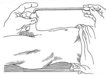
The current treatment methods for urethral stricture mainly include: urethral dilation, urethral endotomy, laser therapy, urethral stent, open urethral reconstruction including urethral end-to-end anastomosis and substitute urethral reconstruction, etc. The selection of treatment methods should be based on the cause, location, length, complications, and previous surgical history of each patient to formulate an individualized treatment plan.
The content of this article is sourced from the internet. The author assumes no responsibility for it. Without permission, copying is strictly prohibited. |

浙公网安备33021202001967号